Learn vocabulary terms and more with flashcards games and other study tools.
Popliteal fossa floor oblique popliteal ligament.
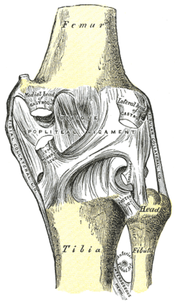
It is attached above to the upper margin of the intercondyloid fossa and posterior surface of the femur close to the articular margins of the condyles and.
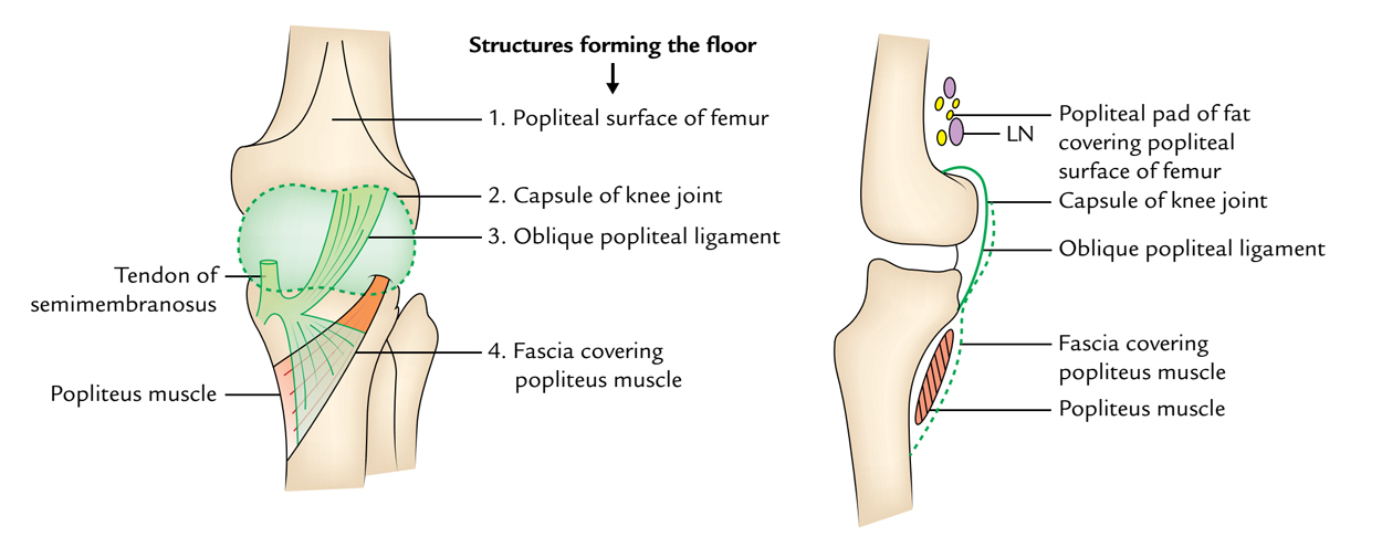
Floor of the popliteal fossa i e popliteal surface of femur posterior aspect of the knee joint.
The popliteal fascia the roof of the popliteal fossa is tough and non extensible and so an aneurysm of the popliteal artery has consequences for the other contents of the popliteal fossa.
The floor of popliteal fossa obliquely from medial to lateral side to reach the lower border of the popliteus muscle where it terminates by dividing into.
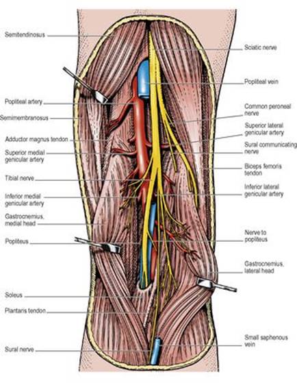
Structures within the popliteal fossa include from superficial to deep.
Floor or anterior wall.
Anterior posterior tibial arteries.
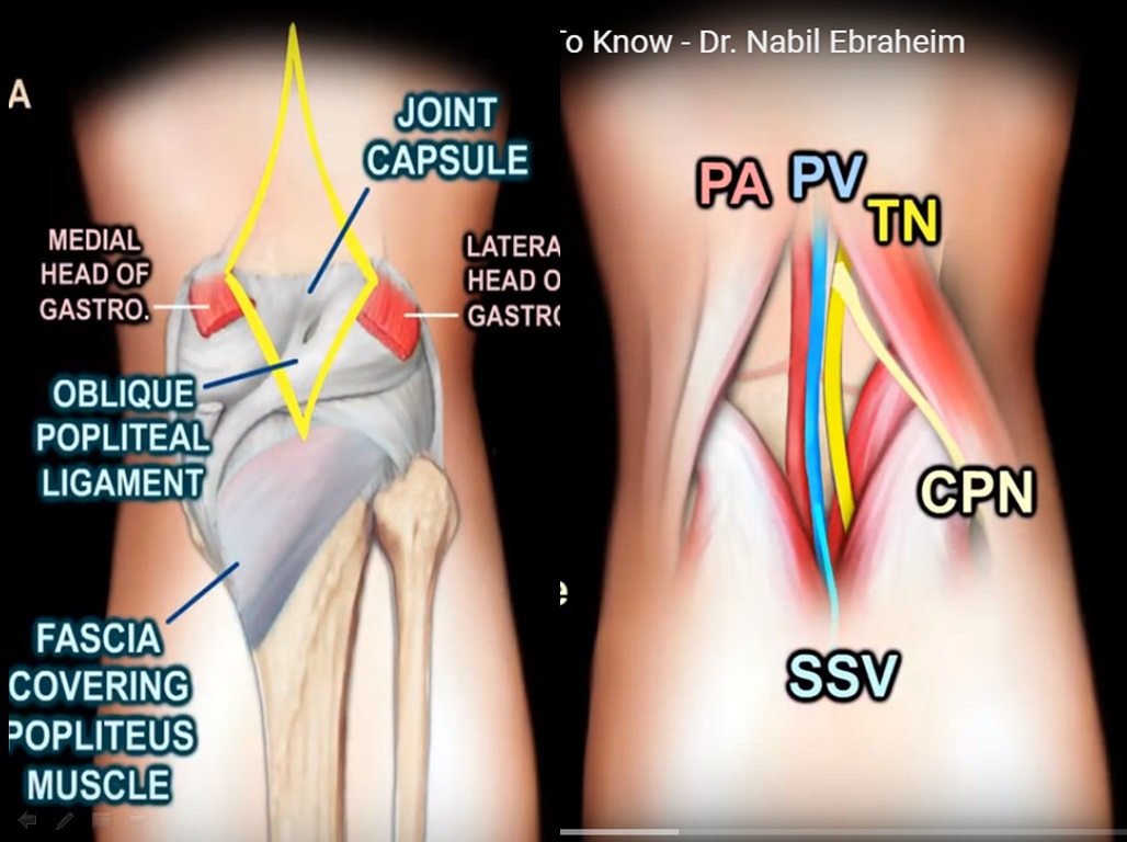
The capsule of the knee joint and oblique popliteal ligament.
The oblique popliteal ligament posterior ligament is a broad flat fibrous band formed of fasciculi separated from one another by apertures for the passage of vessels and nerves.
The popliteal fascia covering the popliteus muscle.
The popliteal fossa sometimes referred to as the hough 1 or kneepit in analogy to the armpit is a shallow depression located at the back of the knee joint the bones of the popliteal fossa are the femur and the tibia like other flexion surfaces of large joints groin armpit cubital fossa and essentially the anterior part of the neck it is an area where blood vessels and nerves pass.
The oblique popliteal ligament posterior ligament is a broad flat fibrous band formed of fasciculi separated from one another by apertures for the passage of vessels and nerves.
The floor is formed by.
The capsule of the knee joint and the oblique popliteal ligament.
The tibial nerve is particularly susceptible to compression from the popliteal artery.
It s created from above downward by.
Learn vocabulary terms and more with flashcards games and other study tools.
It is attached above to the upper margin of the intercondyloid fossa and posterior surface of the femur close to the articular margins of the condyles and below to the posterior margin of the head of the tibia.
Start studying popliteal fossa knee joint.
Popliteal artery a continuation of the femoral artery.
Start studying popliteal fossa.
The popliteal surface of the femur.
The popliteal surface of the femur.